Acoustic neuroma (cerebellopontine angle tumor)

hearing loss
Tinnitus
Dizziness and balance disorders
Nystagmus
Pain and sensory disturbance in the face
occipital headache
Difficulty swallowing
High frequency hearing loss
Mutation in the Schwann cells
surgical procedure
Radiotherapy

Basics

An acoustic neuroma is a rare, benign tumor of the vestibulocochlear nerve. The vestibulocochlear nerve is the 8th of a total of 12 cranial nerves and transmits signals for the sense of hearing and balance. The acoustic neuroma usually arises from Schwann cells of the vestibular part of the vestibulocochlear nerve, which is why the medical name for the tumor is also vestibular schwannoma.

The 8th cranial nerve: vestibulocochlear nerve

Vestibular schwannomas form from uncontrolled growth of Schwann cells in the nerve sheath of the vestibulocochlear nerve. The nerve is a so-called twin nerve, i.e. it has two different parts. One part (auditory nerve, cochlear nerve) is responsible for the sense of hearing, while the other part (vestibular nerve) is important for the sense of balance. Both nerves emerge close to each other in the brain stem and run via the cerebellopontine angle to the inner ear. As the acoustic neuroma usually forms from the Schwann cells of the vestibular nerve, the tumor can either grow laterally on the vestibular nerve or separate the two parts of the mixed nerve, so to speak. The type of tumor growth has a decisive influence on the type of therapy and the prognosis.

Slow growth in the cerebellopontine angle

The tumor grows in the internal auditory canal and often slowly expands from there into the cerebellopontine angle. This is why the tumor is also known as cerebellopontine angle tumor. If the tumor is still located in the bony auditory canal, its location is also referred to as "intrameatal". If the tumor grows into the cerebellopontine angle - a space to the right and left of the brain stem - its location is also referred to as "extrameatal". In most cases, this expanding growth leads to increasing pressure on the vestibular nerve and the cochlear nerve. As the disease progresses, the trigeminal nerve (5th cranial nerve) and the facial nerve (7th cranial nerve) can also be affected. Symptoms that can occur in the course of an acoustic neuroma include unilateral hearing loss, dizziness, tinnitus and paralysis of the facial nerve. The function of other cranial nerves and the brain stem can also be impaired.

Schwerhörigkeit (iStock / eternalcreative)

Incidence

The incidence of acoustic neuroma in Western Europe is around 1 in 100,000, with people between the ages of 30 and 50 being most frequently affected. This means that around 8 to 10 out of every 1,000,000 inhabitants are diagnosed with an acoustic neuroma. Acoustic neuromas account for around 7 % of all brain tumors and over 80 % of all tumors in the cerebellopontine angle. However, it is assumed that many small acoustic neuromas remain undetected and thus unregistered due to a lack of symptoms. The most common symptom in those affected is hearing loss.

Causes

The exact causes of an acoustic neuroma are still unknown. In most cases, the tumor forms from the so-called Schwann cells, which are located around the vestibular nerve and insulate the nerve electrically. This insulation significantly increases the speed of conduction in the nerve. This contributes to faster transmission of stimuli to the sense of balance or hearing.

Non-hereditary tumor

An acoustic neuroma is currently considered a non-hereditary tumor. In rare cases, however, acoustic neuroma can also occur in connection with the hereditary disease neurofibromatosis type 2. If an acoustic neuroma occurs on both sides at the same time (around 5% of cases), this is a strong indication of neurofibromatosis type 2. The disease should then be ruled out or confirmed with the help of genetic tests.

Symptoms

The symptoms of an acoustic neuroma can be divided into early and late symptoms. However, it is not the tumor itself that is responsible for the symptoms, but the impairment of the surrounding structures. An acoustic neuroma can therefore remain symptom-free and unnoticed for a long time. Complaints often only arise when the tumor displaces other structures, disrupting their function.

Early symptoms

As acoustic neuromas usually grow slowly, they generally cause a gradual onset of symptoms. The first symptom in over 94% of those affected is increasing, one-sided hearing loss. This hearing impairment mainly affects the higher tones (high-frequency hearing loss). Bilateral hearing loss occurs in around 5% of cases. This finding is indicative of a genetic disease (neurofibromatosis type 2). Around 80 % of all those affected report tinnitus in addition to high-frequency hearing loss. Other early symptoms of an acoustic neuroma are unsteady gait, vertigo, spontaneous nystagmus and sudden deafness.

Late symptoms

Typical late symptoms of an acoustic neuroma are

  • Peripheral facial nerve palsy (7th cranial nerve)

  • A sensory disorder (hypesthesia) in the area of the 1st and 2nd trigeminal branch (5th cranial nerve)

  • Swallowing disorders

  • Other symptoms due to the compression of cranial nerves and the brain stem

As the tumor is located in the cerebellopontine angle, cerebellopontine angle syndrome sometimes occurs as the tumor grows. Cerebellopontine angle syndrome refers to a combination of symptoms that can occur when there is a mass in the area of the cerebellopontine angle.

Reduction in hearing ability

As described, the tumor usually develops from the Schwann cells of the vestibular nerve. If fibers of the cochlear nerve (auditory nerve) are irritated or compressed by the tumor, this often leads to increasing hearing loss on the affected side. Those affected usually first notice a hearing loss in the high frequencies (e.g. birdsong). This hearing loss can also be an incidental finding during an ENT examination. As the tumor grows, nerve fibres of the vestibular nerve (balance nerve) are usually impaired in their function, causing dizziness in those affected.

Tinnitus

In addition to a reduction in hearing, tinnitus can also occur as a result of the disease. This is a disturbing or unpleasant sound that is constantly heard and has no trigger in the environment. For most people affected, tinnitus is more disturbing and noticeable than the mild hearing loss that usually occurs at the beginning of acoustic neuroma.

Restriction of the organ of balance

As the tumor grows, the restrictions become increasingly severe - dizziness and balance disorders often occur in those affected. Both spinning and swaying dizziness can occur. These symptoms can severely affect the daily lives of those affected. In addition to dizziness symptoms, so-called nystagmus can also occur in the course of an acoustic neuroma. Nystagmus describes an involuntary, rhythmic-periodic movement of the eye, for example when tracking or fixating objects in the environment.

Intracranial pressure in large acoustic neuromas

If a vestibular schwannoma remains undetected for a long time, it can also lead to so-called intracranial pressure symptoms. These occur when the pressure in the skull increases due to the growing tumor.

Common symptoms are headaches, which occur mainly in the back of the head, visual disturbances or even loss of vision, vomiting and changes in personality. Symptoms of intracranial pressure should always be clarified and treated immediately by a doctor.

Diagnosis

The diagnosis of an acoustic neuroma begins with a detailed medical history taken by the attending physician. Here, the patient is questioned in detail about the symptoms and the time of occurrence. The medical history can already provide important information for the treating physician. The suspicion of an acoustic neuroma is then usually confirmed by further examinations. ENT specialists or neurologists are often involved in these examinations.

Hörtest (iStock / DuxX)

Hearing test

A hearing test is one of the first tests to confirm a suspected acoustic neuroma, as the disease often manifests itself in reduced hearing. Sound threshold audiometry often reveals a so-called high-frequency hearing loss. However, as those affected can state what they hear in the test themselves, it is always a subjective test. Audiometry can be used to determine which ear is hearing impaired.

Checking the balance

Initial indications of damage to the sense of balance can also be obtained through simple tests. For example, those affected have to perform certain walking and standing tests with their eyes closed. The Romberg and Unterberger tests in particular provide important indications of damage to the vestibular system. An existing nystagmus (twitching of the eyes) is also an indication of a restriction of the sense of balance.

Brainstem audiometry

Brainstem evoked response audiometry (BERA) can also be carried out to investigate an acoustic neuroma. This involves testing the auditory nerve using a loudspeaker. Various click sounds are played to the patient, which are measured via an electrode behind the ear without the patient's active participation. The information from the sound is transmitted directly to the brain via the auditory nerve and recorded by measurement sensors.

MRT-Untersuchung (iStock / shironosov)

Radiological examinations

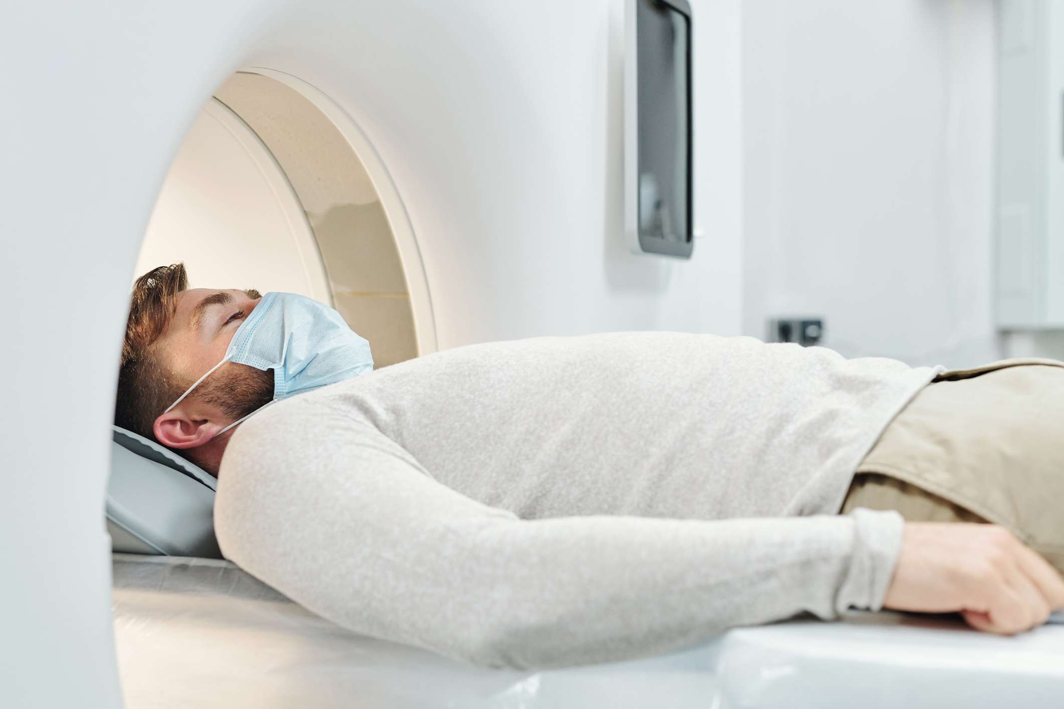
The suspected diagnosis of an acoustic neuroma is usually confirmed with the help of an imaging examination. Computed tomography (CT) and magnetic resonance imaging (MRI) play an important role here.

MRI is particularly important for early detection, as it does not cause any radiation exposure for those being examined. With the help of this procedure, neurinomas that are only a few millimetres in size can be detected by administering a contrast medium.

Therapy

Depending on the growth and extent of the tumor, there are three possible strategies for treating acoustic neuroma.

Wait and see

Small acoustic neuromas with a diameter of only a few millimeters that do not cause any symptoms can often be observed. As the growth of most acoustic neuromas is very slow, surgery or radiotherapy can at least be delayed. In older patients, the tumor often grows at an even slower rate.

Surgical removal

If the tumor exceeds a size of around 3 centimetres in diameter, the tumor should be surgically removed, as healthy tissue is inevitably irradiated during radiotherapy. Surgical removal is carried out while sparing the surrounding structures (healthy tissue, blood vessels and cranial nerves), which can be well preserved in most cases.

Radiotherapy

As an alternative to surgery, smaller tumors can be treated with radiation using a gamma or cyber knife. The advantage of radiotherapy is that patients can avoid the potential risks of surgery. During treatment, the cells of the tumor are destroyed or altered by radiation. The Cyber-Knife is a robot-assisted linear accelerator for the treatment of brain tumors.

Forecast

Acoustic neuromas are characterized by the fact that they generally grow very slowly and do not form metastases. The tumors correspond to WHO grade 1 in the classification of brain tumors. The mortality rate for acoustic neuromas is well below 1% of those affected. In around 95% of cases, the cranial nerves can be preserved during treatment. For tumors with an average diameter of less than 3 cm, the cranial nerves can even be preserved in almost 100% of treatment cases.

Good prognosis with early diagnosis

As with all other tumors, the earlier the tumor is diagnosed and treated, the better the prognosis. Depending on the size of the tumor, hearing is preserved in around 50-70% of those affected. Larger tumors usually have to be removed with the help of surgical interventions, although they generally do not recur. Exceptions are large tumors that have not been completely removed by surgery and tumors that have not responded or have responded poorly to radiotherapy. These tumors may relapse (recurrence).

Regular check-ups after removal

As some acoustic neuromas tend to recur, it is advisable to have regular MRI checks after tumor removal. This allows even very small recurrences to be detected, which can then be treated surgically or with radiotherapy. However, small and medium-sized tumors in particular can often be removed without complications.

Dr. med. univ. Moritz Wieser

Dr. med. univ. Moritz Wieser

Thomas Hofko

Thomas Hofko


Logo

Your personal medication assistant

Drugs

Browse our extensive database of medications from A-Z, including effects, side effects, and dosage.

Substances

All active ingredients with their effects, applications, and side effects, as well as the medications they are contained in.

Diseases

Symptoms, causes, and treatments for common diseases and injuries.

Social media

The presented content does not replace the original package insert of the medication, especially regarding the dosage and effects of individual products. We cannot assume liability for the accuracy of the data, as the data has been partially converted automatically. Always consult a doctor for diagnoses and other health-related questions.

© medikamio