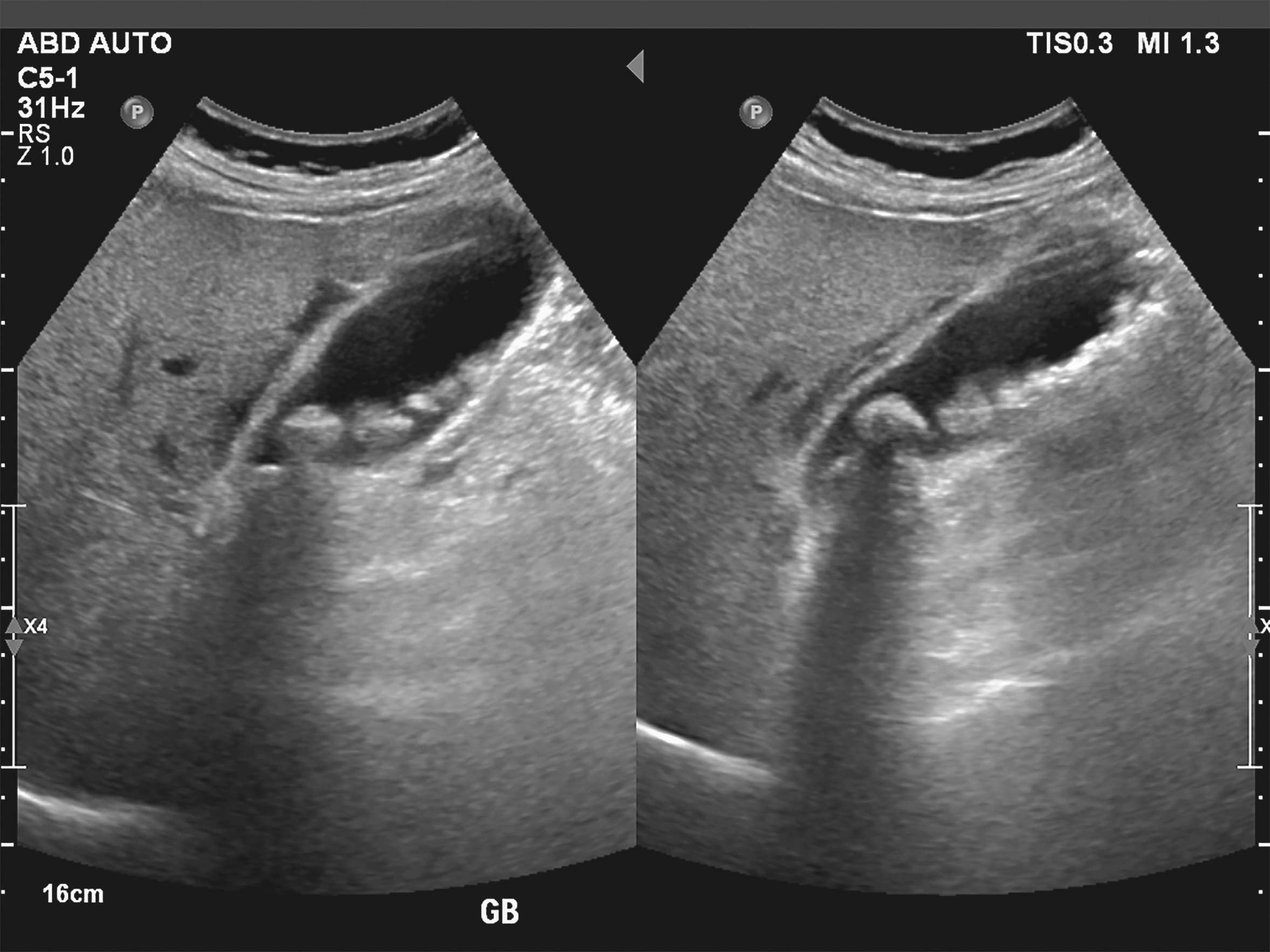
Gallstones (cholelithiasis)

yellowing of the skin
upper abdomen pain
Biliary colic
Pain during food intake
Feeling of fullness
Nausea
Vomit
dark urine
Chills
Fever
bile deposition
Supersaturation of the bile with cholesterol
female, overweight, several children, forty, light-skinned/light hair
High age
Pregnancy
Overweight
positive family history
diabetes mellitus
Rapid weight reduction
Disturbed enterohepatic circulation
Medication
Antibiotics
surgical procedure

Basics

The term cholelithiasis refers to the presence of gallstones. These can occur either in the gallbladder (cholecystolithiasis), the bile ducts (choledocholithiasis) or in the small bile ducts of the liver (cholangiolithiasis). Around 10 to 15 % of the population have gallstones, although most of those affected remain symptom-free. Small gallstones are also often referred to as bile sand, sludge or gallstones.

The human body produces around 700 ml of yellow-brown to olive-green bile per day, which enables (fat) digestion in the small intestine. This consists of

  • Water (82 %)

  • electrolytes

  • Bile acids (subject to the enterohepatic circulation)

  • Phospholipids (especially lecithin)

  • cholesterol

  • bilirubin

  • proteins

The most common forms of gallstones are cholesterol, pigment and mixed gallstones. Cholesterol stones and mixed gallstones together account for around 80 % of gallstones in industrialized countries. Pigment stones account for around 20% of gallstones.
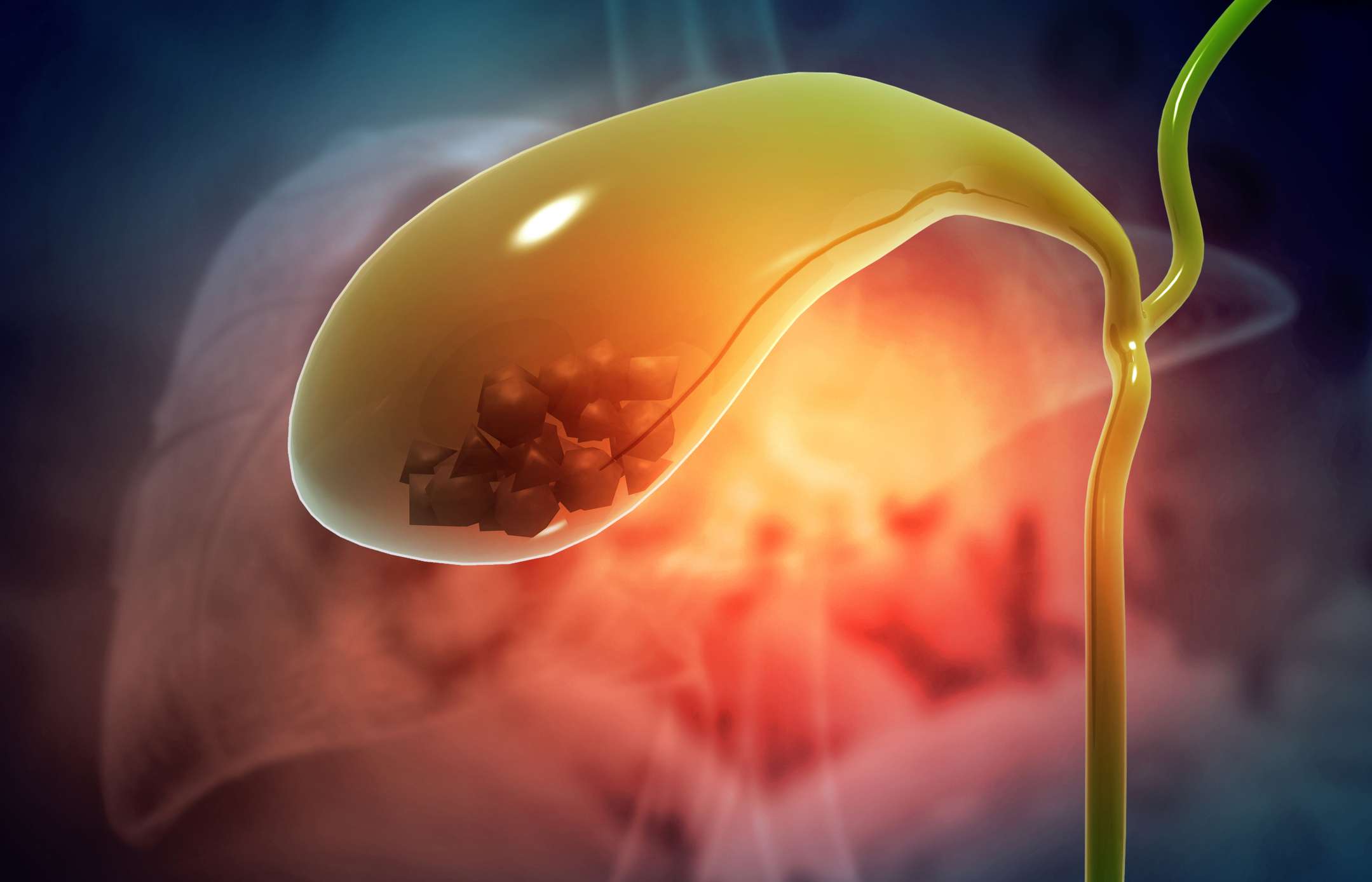
Gallensteine (iStock / Rasi Bhadramani)

Cholesterol stones, pigment stones and mixed gallstones

Cholesterol stones often form when the ratio of lecithin, cholesterol and bile acids within the bile is out of balance. This often leads to supersaturation of the bile with cholesterol and subsequent stone formation (lithogenesis). An additional factor in the formation of cholesterol stones is reduced gallbladder movement (gallbladder motility). Mixed gallstones often consist of cholesterol and pigment or calcium salts. Their cholesterol content is usually over 70 %. Pigment gallstones consist mainly of bilirubin and bilrubin degradation products. Their cholesterol content is often less than 20 %.

Frequency

The incidence of gallstones in Europe is 9% in women and 5% in men. The risk of developing gallstones increases steadily with age. First-degree relatives of people with gallstone disease have a 4.5-fold increased risk of also developing gallstones.

Causes

Gallstones are caused by an incorrect composition of the bile. If there is an imbalance of soluble substances, gallstones can form.

Risk factors for the development of gallstones:

  • older age

  • female gender

  • pregnancy

  • beingoverweight (obesity)

  • positive family history

  • certain ethnic background

  • Diabetes (diabetes mellitus type 2)

  • Rapid weight reduction (e.g. dieting)

The most important risk factors for gallstones can be easily remembered using the 6F rule. These are: female, fat, fertile, forty, familial and fair.

Underlying diseases that can favor the occurrence of gallstone disease are

  • a disturbed enterohepatic circulation (e.g. in Crohn's disease)

  • Haemolytic anaemia

  • hyperparathyroidism

  • LPAC syndrome ("low phospholipid associated cholelithiasis")

  • Cystic fibrosis

  • Meulengracht's disease

  • Myotonic dystrophy

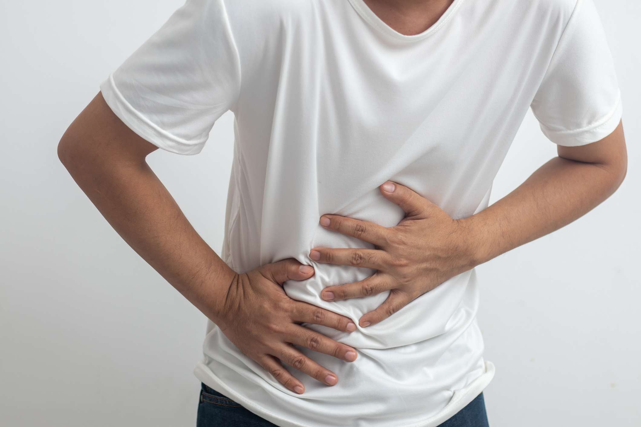
Oberbauchschmerzen (iStock / Vachiravit Vasuponsritara)

Cholecystitis

Cholecystitis describes a bacterial inflammation of the gallbladder, which is usually caused by the pathogens Escherichia coli, Klebsiella, Enterobacter or anaerobic bacteria. Acute calculous cholecystitis occurs in 90% of cases due to stone formation with stasis, obstruction and microtrauma of the gallbladder wall. Serious illnesses, operations or trauma are often the triggers of acalculous cholecystitis (so-called stress gallbladder). Chronic cholecystitis is a secondary condition of repeated, acute gallbladder inflammation. Extreme forms are the shrunken gallbladder (scarred atrophy) and the porcelain gallbladder (scarred calcification).

Cholangitis

Stones or scarring promote inflammation of the bile ducts (cholangitis). The causative, ascending bacteria usually originate from the small intestine. Endoscopic retrograde cholangiopancreatography (ERCP) is also a risk factor for cholangitis.

Symptoms

Gallstones often cause no symptoms, so that around 75% of those affected remain asymptomatic. The typical symptom of gallstones in the gallbladder (cholecystolithiasis) is biliary colic. Symptoms are also frequently triggered by an obstruction or inflammation of the bile ducts.

Patients with symptomatic gallstones (around 25% of those affected) have an increased risk of a number of complications (e.g. pancreatitis, gallbladder inflammation).

Symptoms that can occur as a result of gallbladder disease are

  • Cramp-like or persistent pain in the right upper abdomen (colic)

  • Dull, non-specific upper abdominal pain

  • Pain after eating

  • Pain that radiates to the right shoulder

  • Feeling of fullness

  • Nausea

  • vomiting

If the stones are located in the bile ducts and obstruct the outflow of bile, further symptoms may occur:

Biliary colic lasting more than 5 hours is usually caused by a complicated gallstone disease. About half of all people with biliary colic experience another colic or other gallstone complications within a year.

Diagnosis

In the case of symptomatic gallstone disease, a detailed medical history and clinical examination findings are crucial for the diagnosis. Pressure pain often occurs in the right upper abdomen.

An ultrasound examination of the upper abdomen (upper abdominal sonography) is particularly suitable for diagnosing gallstones, for example in the gallbladder. Upper abdominal sonography can also be used to assess the intrahepatic bile ducts. Computed tomography (CT) is particularly suitable in complicated cases or in an emergency situation - here, for example, a gallbladder perforation or pancreatitis can be detected.

The Murphy sign describes an examination in which the patient takes a deep breath and the doctor simultaneously palpates with his fingers under the right costal arch (area of the gallbladder). If the gallbladder is inflamed, this causes pain and the doctor can feel the gallbladder.

Laboratory examination

The blood test is usually unremarkable in the case of simple biliary colic. In the case of gallbladder inflammation or inflammation of the bile ducts, there is often an increase in white blood cells (leukocytosis) and an increase in C-reactive protein (CRP).

Gallenblasensonographie (iStock / Shidlovski)

Further diagnostics

Another method for diagnosing gallstones is endoscopic cholangiography (ERCP). With this method, smaller stones can also be removed during the examination. Alternatively, magnetic resonance cholangiography (MRCP) can also be used. With this method, the area surrounding the bile ducts is also visualized at the same time. Endosonography is also suitable as an imaging procedure.

Therapy

Asymptomatic gallstone disease does not need to be treated. Only around 1% of asymptomatic gallstones develop into gallstones with symptoms within a year. However, if symptoms occur in the course of the gallstones, this is usually an indication for laparoscopic removal of the gallbladder (cholecystectomy). The recurrence rate for symptomatic gallstone disease is around 50% with a complication rate of 1 to 3% per year.

Symptomatic therapy

Spasmolytics (e.g. intravenous N-butylscopolamine) can be used for acute biliary colic. Contraindications such as glaucoma, cardiac arrhythmia or pregnancy should be taken into account. Painkillers such as metamizole, paracetamol or opiate derivatives such as pethidine are also suitable for the symptomatic treatment of pain. Antibiotic therapy (e.g. ceftriaxone plus metronidazole) is often recommended for acute gallbladder inflammation or inflammation of the bile ducts (cholangitis). The most common pathogens of gallbladder inflammation are enterococci or Escherichia coli.

Conservative stone therapy

If the risk of surgery is high, conservative litholysis using ursodeoxycholic acid (UDCA) can be performed. However, this is only recommended for small gallbladder stones < 5 mm. In addition, therapy should be continued for a further three months if the patient is stone-free (confirmed by ultrasound).

Forecast

Complications that can occur in the course of gallstone disease are

  • Acute inflammation of the gallbladder(cholecystitis)

  • Inflammation of the bile ducts (cholangitis)

  • Inflammation of the pancreas(pancreatitis)

  • Jaundice (icterus)

  • Increased risk of gallbladder or bile duct cancer

Operations on the gallbladder and bile ducts are relatively low-risk routine procedures and are therefore generally well tolerated by patients. In addition, the complication rate is usually low. Unfortunately, non-surgical treatments for gallstone disease have a high recurrence rate.

The most common complication of gallstone disease is acute inflammation of the gallbladder (cholecystitis) due to obstruction of the bile duct. In rare cases, the stone may also break through into the abdominal cavity or the small intestine (perforation). In most people, the bile duct leads into the small intestine together with the pancreatic duct. An obstruction with a backlog can therefore trigger an inflammation of the pancreas(acute pancreatitis).

Prevent

No specific measures are currently known to prevent gallstones. In general, a healthy lifestyle with a balanced diet and regular physical activity (sport) is recommended.

Risk factors for gallstones are obesity and a diet high in cholesterol and low in fiber. These factors should be kept as low as possible, even if gallstones have already occurred in the past.

Dr. med. univ. Moritz Wieser

Dr. med. univ. Moritz Wieser


Logo

Your personal medication assistant

Drugs

Browse our extensive database of medications from A-Z, including effects, side effects, and dosage.

Substances

All active ingredients with their effects, applications, and side effects, as well as the medications they are contained in.

Diseases

Symptoms, causes, and treatments for common diseases and injuries.

Social media

The presented content does not replace the original package insert of the medication, especially regarding the dosage and effects of individual products. We cannot assume liability for the accuracy of the data, as the data has been partially converted automatically. Always consult a doctor for diagnoses and other health-related questions.

© medikamio