Urethral stricture

Urethral stricture
International Classification (ICD) N35.-
Symptoms weakened urine stream, prolonged bladder emptying time, nocturnal leakage of urine (nocturia), Residual urine formation, Feeling of incomplete bladder emptying, increased urination (pollakiuria), painful emptying of the bladder (dysuria), Macrohematuria, Urinary tract infections, Difficulty emptying the urinary bladder
Possible causes Genitourinary disease
Possible risk factors surgical interventions, previous urinary tract infections, Without apparent cause (idiopathic)
Possible therapies surgical procedure, Dilation (Dilatation), Open reconstruction

Basics

The term urethral stricture refers to a narrowing of the urethra. The medical term is derived from the Latin word "strictura", (engl.: to contract).

About 1% of the population suffers from urethral stricture, and about half of all cases are either medically caused or occur due to unknown causes (idiopathic). The incidence of urethral stricture increases significantly with age, rising to over 100 cases per 100,000 population in people over 65 years of age. It mainly affects men.

Depending on the severity and duration of symptoms, urethral stenosis can have long-term negative effects on the entire urinary tract. Urethral strictures cause continuous damage to the entire urinary tract due to functional obstruction. They can also lead to reduced kidney function if they progress chronically.

Although it is a common urological condition, there are no guidelines on urethral stricture from the European Association of Urology (EAU). Various treatment options are available depending on the anatomical location of the stricture. The treatment options for urethral stricture should always be selected after education and diagnosis by the treating physician. Likewise, the individual preferences of the affected person play a major role in the treatment decision.

Urethral stricture in women

In contrast to men, urethral stricture rarely occurs in women. In women, traumatic (e.g. after childbirth) or medically induced urethral strictures (e.g. after radiation) are usually in the foreground. However, the pathogenesis of this disease is not yet fully understood.
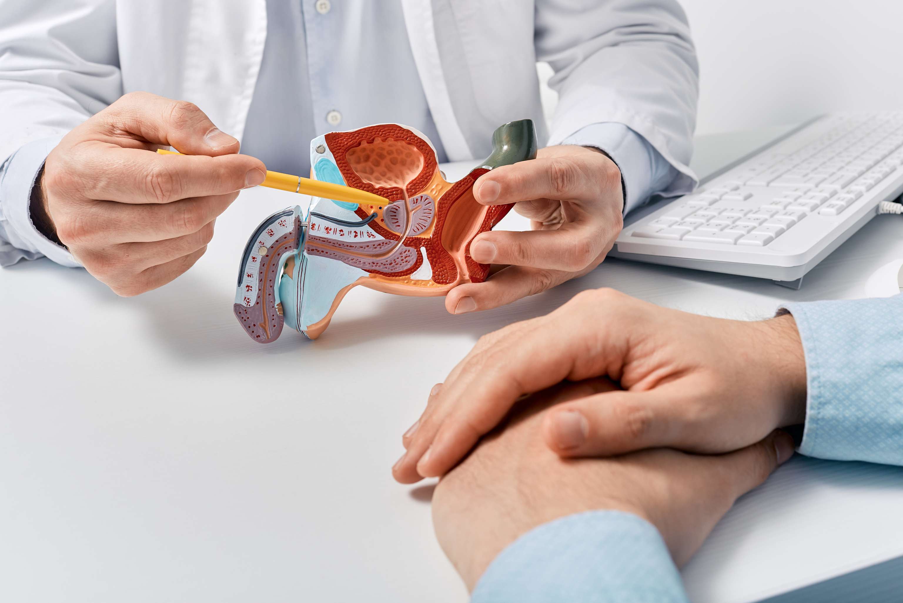
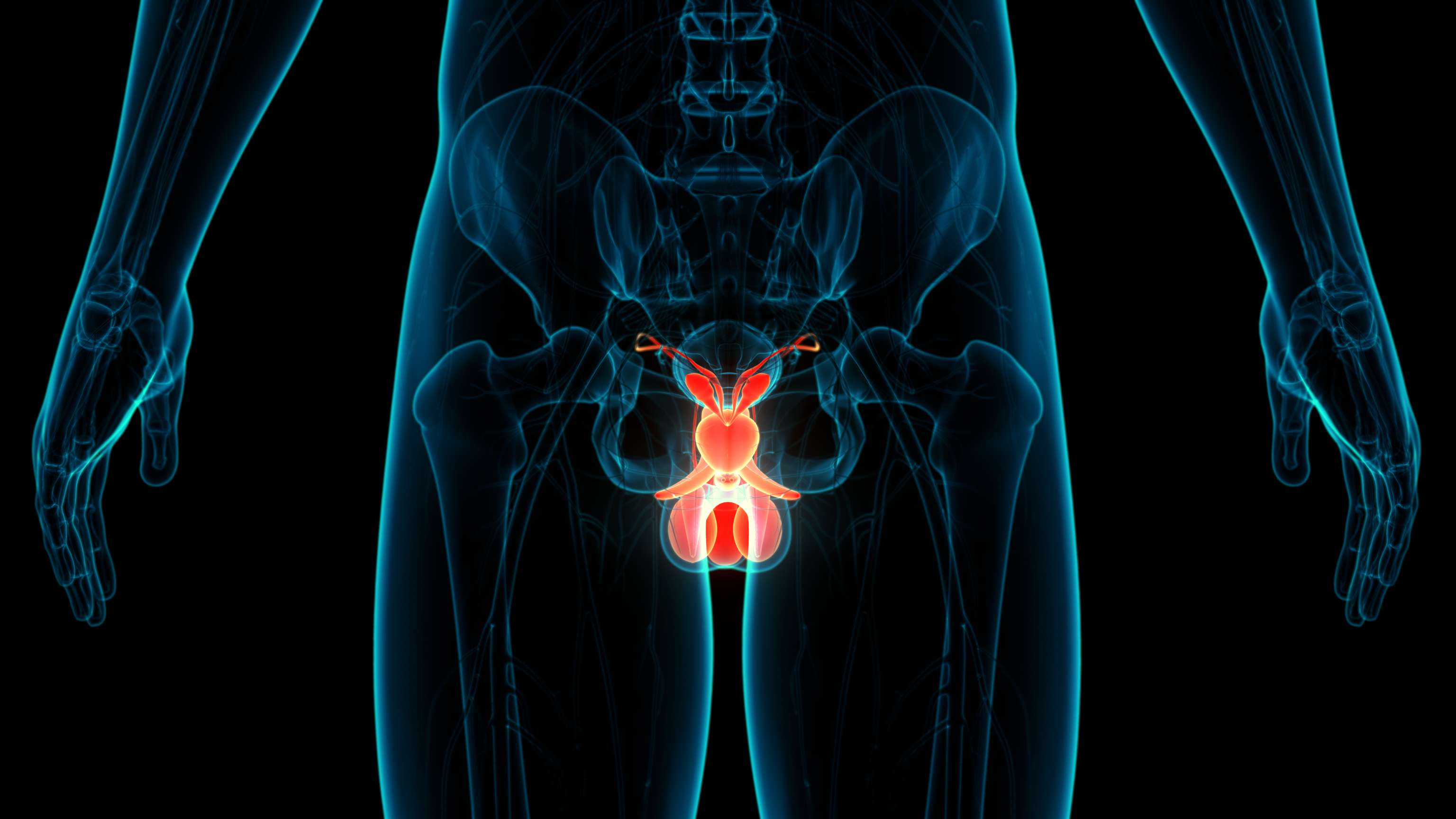
The male urogenital tract Der männliche Urogenitaltrakt (iStock / peakSTOCK)

Classification of urethral stricture

The urethra can be divided into anterior (anterior) and posterior (posterior) sections. The prostatic and membranous urethra belong to the posterior urethral section. Consequently, narrowings in this area are also called posterior urethral strictures. Narrowings in the region of the penile root and penis, however, are much more common and belong to the anterior urethral strictures. Depending on their location, these are classified as bulbar, penile, and glandular urethral strictures, as well as strictures of the meatus urethrae externus.

After accurate diagnosis, urethral strictures can be divided into short-stretch (≤1 cm) and long-stretch (>1 cm) strictures.

The bulbar urethra is most commonly affected by urethral stricture (approximately 50% of all cases). It has the most favorable prognosis. The reason for this is the good tissue perfusion due to the strongly formed and surrounding urethral corpus spongiosum.


Causes

In most cases, urethral strictures develop on their own (idiopathic) or are caused medically (iatrogenic). Medical causes often include surgery or even manipulation of the urethra. Examples include indwelling catheters, cystoscopy, or surgeries such as radical prostatectomy or transurethral resection. Urethral stenosis can also occur as a late complication. Traumatic causes are also a common cause of urethral stricture (e.g., after pelvic trauma). Other causes of urethral stricture are infectious urinary tract infections. Since nowadays antibiotics are usually prescribed for the therapy of urinary tract infections, post-traumatic and physician-induced urethral strictures - compared to urethral strictures after inflammations - are gaining in importance.

The urogenital system Das Urogenitalsystem (iStock / magicmine)

Histological changes in urethral stricture

The causes described above lead to scarring and fibrosis of the tissue lining the urethra, resulting in narrowing of the lumen.

Symptoms

Complaints that are common with urethral stricture are:

  • A weakened urinary stream

  • A prolonged bladder emptying time

  • Nighttime leakage of urine (nocturia)

  • Residual urine formation

  • Feeling of incomplete bladder emptying

  • Increased urge to urinate (pollakiuria)

  • Painful emptying of the bladder (dysuria)

  • Urinary retention up to kidney congestion

When first manifesting, affected individuals usually have frequent urinary retention, chronic retention bladder, visible blood in the urine (macrohematuria), or recurrent urinary tract infections. If urethral stricture occurs in the lower part of the urethra, it may also cause a rotated or divergent urinary stream (palmuria). The divergent urine stream can interfere with bladder emptying and reduce the quality of life. In very severe cases, urine may back up into the kidneys with subsequent renal insufficiency.

Diagnosis

An exact diagnostic clarification is necessary to select the ideal therapeutic path for the treatment of urethral strictures. Several examinations are available for this purpose.

As a rule, a urine flow measurement (uroflow) is performed, in which a possible weakening of the urinary stream can be diagnosed by means of a special device. By means of an ultrasound examination, a sonographic residual urine measurement can be performed. Classical diagnostic procedures are retrograde urethrography and micturition cysturethrography, in which the urethra is visualized by means of an X-ray image after prior application of a contrast medium.

Another method is cystoscopy, in which the urethra and urinary bladder are examined with an endoscope. In most cases, a urine examination is also performed to detect possible blood impurities.

Therapy

If these conservative methods are not possible or not sufficient, surgical therapy is chosen. In an operation, for example, the narrowed segment can be removed and the urethral segments before and after it sutured (end-to-end anastomosis) or the resulting gap can be bridged with the help of other mucosal tissue, for example from the mouth.

In particular, the treatment of patients with multiple stricture recurrences is often challenging. In any case, the right therapy option should be selected individually and according to the wishes of the patient.

Blasenkatheter (iStock / Iuliia Alekseeva)

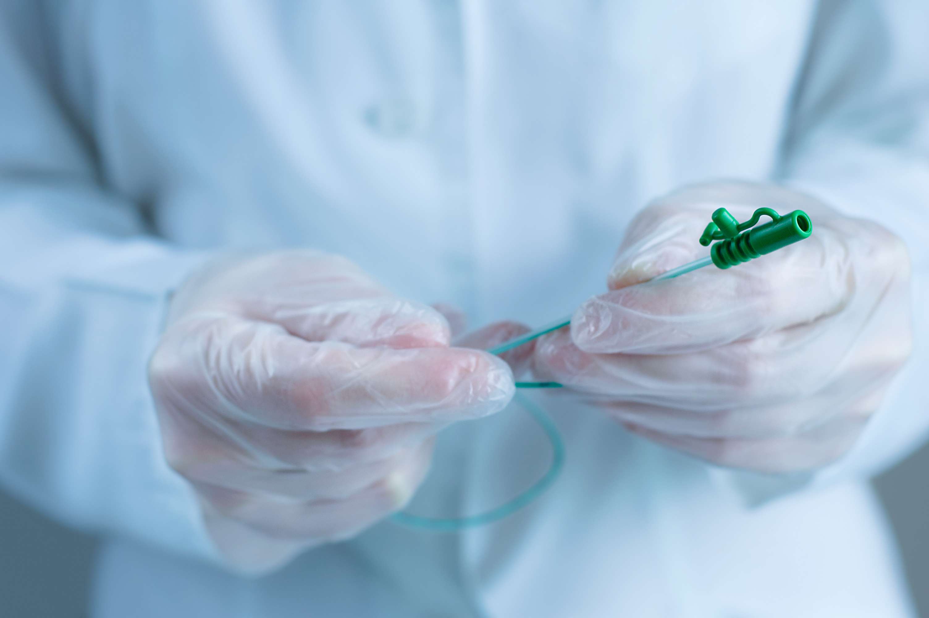
The therapeutic options in the treatment of urethral stricture - regardless of the anatomical localization of the urethral stricture - are diverse. The options include bougienage, dilatation, internal urethrotomy (urethral slit) or open reconstruction.

In bougienage, the narrowed area is mechanically dilated using a surgical instrument (bougienage pin). Dilation works similarly, usually using a balloon catheter to dilate the urethra. Another option is to cut open the stricture as part of a urethral slit (urethrotomy). A major disadvantage of these methods is the high risk of recurrence of up to 80%. For this reason, drug-coated balloon catheters have been available for several years. Here, the chemotherapeutic agent paclitaxel is used to prevent the treated urethra from scarring again.

Forecast

Urethral stricture is a common disease that mostly affects men. Especially due to the high recurrence rate after conservative therapy attempts and the often protracted clinical courses, the quality of life of those affected is often severely impaired. Surgical therapy methods often produce good results, but have risks and require specialized urologists. New drug-eluting catheter dilatations have shown good results in studies. However, dissemination and scientific evidence is still limited.

Editorial principles

All information used for the content comes from verified sources (recognised institutions, experts, studies by renowned universities). We attach great importance to the qualification of the authors and the scientific background of the information. Thus, we ensure that our research is based on scientific findings.
Dr. med. univ. Moritz Wieser

Dr. med. univ. Moritz Wieser
Author

Moritz Wieser graduated in human medicine in Vienna and is currently studying dentistry. He primarily writes articles on the most common diseases. He is particularly interested in the topics of ophthalmology, internal medicine and dentistry.

Dr. med. univ. Bernhard Peuker, MSc

Dr. med. univ. Bernhard Peuker, MSc
Lector

Bernhard Peuker is a lecturer and medical advisor at Medikamio and works as a physician in Vienna. In his work, he incorporates his clinical knowledge, practical experience and scientific passion.

The content of this page is an automated and high-quality translation from DeepL. You can find the original content in German here.

Ad

Ad

Your personal medicine assistent

afgis-Qualitätslogo mit Ablauf Jahr/Monat: Mit einem Klick auf das Logo öffnet sich ein neues Bildschirmfenster mit Informationen über medikamio GmbH & Co KG und sein/ihr Internet-Angebot: medikamio.com/ This website is certified by Health On the Net Foundation. Click to verify.
Drugs

Search our database for drugs, sorted from A-Z with their effects and ingredients.

Substances

All substances with their common uses, chemical components and medical products which contain them.

Diseases

Causes, symptoms and treatment for the most common diseases and injuries.

The contents shown do not replace the original package insert of the medicinal product, especially with regard to dosage and effect of the individual products. We cannot assume any liability for the correctness of the data, as the data was partly converted automatically. A doctor should always be consulted for diagnoses and other health questions. Further information on this topic can be found here.