1. anamnesis
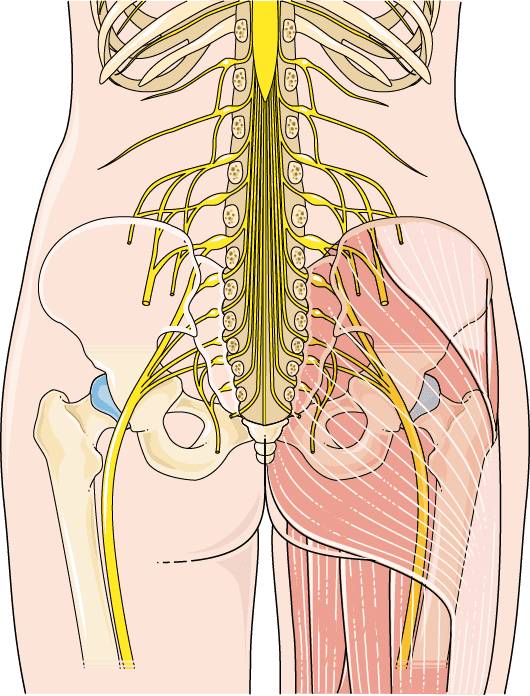
Before a neurological examination is performed, the patient's medical history should first be taken. In this way, a sciatic nerve irritation caused by a herniated disc, for example, can be well diagnosed. Particular emphasis should be placed on body malposition, loss of sensation and signs of paralysis.
2. imaging procedures
X-rays of the spine are taken to obtain a more accurate picture.
To diagnose a herniated disc as the cause, a computed tomography or magnetic resonance imaging is usually performed. In some cases, this can even reveal the location where the herniated disc is pressing on the nerve root. To be on the safe side, a contrast medium image of the spinal canal and the nerve root canals (myelogram or myelo-CT or myelo-MR) can be made.
A rather unusual method today is discography. Here, the contrast medium is administered directly into the area of the intervertebral discs.
3. neurological examinations
An electromyogram (EMG) can be used to determine the location of nerve damage. It can also be used to estimate the duration and extent of nerve damage.
4. blood and nerve fluid diagnostics
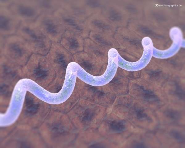
If the treating physician suspects an inflammatory disease or an infection with herpes zoster or Borrelia as the cause, a blood test is usually performed. A spinal fluid (CSF) examination may also be necessary in this case.


